Stereomicroscope-Informed Soundboard Work on Fiber Damage
What the Stereomicroscope Reveals That the Eye Cannot
At 10–40x magnification, a quilt block shifts from a surface-level color problem to a three-dimensional fiber problem. The distinctions become immediately visible. Intact cotton fibers have a twisted ribbon structure with a smooth cuticle; UV-degraded fibers lose that structure, developing microcracks and a matte surface texture that scatters light differently and holds dye differently. Fibers damaged by batting contact show flattening and fiber-to-fiber adhesion from prolonged compression. Wash-damaged zones show fiber tip abrasion and, in fugitive dye cases, a characteristic dye accumulation at fiber intersections where the migration stopped.
Each of these fiber states responds differently to a dye bath. Intact, mordant-receptive fibers will take color at the expected concentration. UV-degraded fibers with compromised cellulose structure will take color inconsistently — some areas oversaturating, others remaining pale — because the mordant bonding sites have been partially destroyed. Severely degraded fibers should not receive wet treatment at all without prior consolidation, because the immersion stress can cause physical fiber loss.
Comparative Studies of Cotton Fabric Changes Under Degradation — MDPI Fibers documents the specific color change and embrittlement patterns in cotton under UV, thermal, and chemical degradation, confirming that fiber morphology and dye uptake capacity shift together — which is why visual color assessment alone is insufficient for treatment planning.
Conserving Amish Quilts for Smithsonian American Art Museum documents a real conservation case in which stereomicroscope examination revealed fiber damage that was not apparent at the macro scale, changing the treatment approach entirely before any dye work began. For Amish solid-color quilts — where the uniformity of the ground dye makes subtle damage patterns even harder to read visually — microscope examination is particularly critical.
Mapping Fiber States to Channel Settings
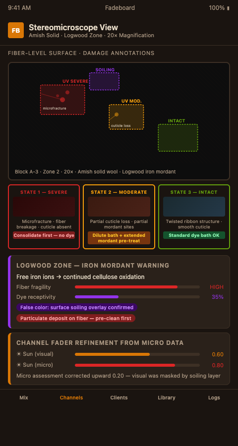
The practical integration of stereomicroscope work into the Fadeboard session runs in two directions. First, the microscope examination refines the channel settings the workshop would have estimated from macro visual inspection alone. Second, the fiber-damage map adds a treatment-feasibility layer that prevents applying dye to zones that cannot safely receive it.
Start with the sun-exposure fader. At macro scale, a visually faded zone suggests a moderate-to-high fader setting. Under the microscope, the same zone might show one of three distinct fiber states: light surface yellowing with intact structure (supports a standard dye bath), moderate microfracturing with partial cuticle loss (supports a diluted dye bath with extended mordant pre-treatment), or severe embrittlement with visible fiber breakage (requires consolidation before any dyeing, or exclusion from dye treatment entirely). The fader setting stays the same — it represents the chromatic damage — but the dye bath formula attached to it diverges based on the fiber assessment.
TSG Chapter V: Fiber Identification Methods — AIC Conservation Wiki specifies polarized light microscopy as the primary tool for textile fiber identification, which is the standard approach when the workshop needs to confirm whether a quilt's degraded fibers are cotton, linen, or a blend — a distinction that affects mordant chemistry and dye bath temperature limits.
Identification of Natural Fibres — Canadian Conservation Institute Notes 13/18 provides the practical guide to microscope-based fiber identification, with reference images that cover the typical calico, chintz, and feedsack cotton varieties found in 19th-century quilts.
The batting-contact fader setting benefits particularly from microscope verification. Batting contact damage is mechanically different from UV damage — it is primarily compression and surface abrasion — and the fiber state under magnification shows that difference clearly. Fibers in a batting-contact zone that have been flattened but not chemically degraded are fully dye-receptive. The fader setting can be translated directly into dye-bath concentration without the dilution correction that UV-damaged fibers require.
Identifying False Color Readings
One of the most practically important functions of stereomicroscope examination is identifying false color readings — zones that appear to be at a specific fade state but are actually presenting a different color due to a physical rather than chemical change.
Surface soiling is the most common false color source. A cotton block that reads as light brown at macro scale may actually be a faded weld yellow overlaid with decades of airborne particulates. Under the microscope, the particulate layer is visible as a discontinuous deposit on the fiber surface rather than an integral color. A dye bath applied to the unexamined surface will produce a muddy, over-saturated result because the workshop is adding dye on top of a layer that can partially lift during wet treatment.
TSG Dye Identification Methods — AIC Conservation Wiki describes methods for linking fiber-level observations to specific dye-failure chemistry, which helps distinguish true fade states from false readings caused by surface contamination, optical interference, or dye migration from adjacent blocks.
The natural-dye fade prediction models that inform forward-looking fader settings are only reliable when the input data — the current chromatic state of each zone — is accurate. Microscope examination is the quality-control step that validates the macro assessment before the session file is finalized.
For logwood black zones in mourning quilts, microscope examination is essential. Logwood black is produced by an iron mordant that causes its own fiber degradation over time — the iron continues to oxidize the cellulose after dyeing, producing a characteristic brittle, tender area that tears easily. Any dye work in a logwood zone without fiber assessment risks accelerating that physical failure.
Advanced Tactics: Integrating Microscope Sessions Into Project Timelines
The logistical question most workshops face is how to fit microscope examination into project timelines without doubling the assessment phase. The answer is selective examination rather than comprehensive coverage.
For a double-wedding-ring quilt with a hundred ring segments, microscope examination of every segment is not practical. The protocol that works is sampling: examine a minimum of three blocks from each identified treatment zone (high-fader, medium-fader, low-fader), plus any block that shows an anomalous macro reading. If the sampled blocks within a zone show consistent fiber state, the whole zone receives the corresponding treatment formula. If the samples diverge, the zone is split.
Microscopes for Paintings and Textiles — ZEISS provides technical guidance on stereo and confocal microscopy setups for textile conservation, including recommendations for documentation photography that integrates with written treatment records. The microscope examination notes become an appendix to the Fadeboard session file — a fiber-state map that complements the channel-setting map.
The restoration dye stability on antique cotton question connects directly to fiber state: a dye applied to severely degraded fibers with poor surface structure will not achieve the expected bond strength, and the resulting color will fade faster than the formula predicts. The microscope examination is therefore not just a damage assessment — it is a reliability check on the predicted dye performance.
The microscope-informed crazing work developed for bisque doll restoration uses the same principle in a different material context: physical surface damage changes the substrate's response to applied pigment, and knowing the damage profile before application is the prerequisite for a predictable result.
Workshops that add systematic stereomicroscope examination to their Fadeboard session protocol before setting channel-based dye formulas are operating at a level that produces consistently better long-term results. If you want to build microscope assessment into your standard workflow, Fadeboard's annotated session files give you the format to record and use what the microscope shows.
Book a Fadeboard consultation and bring your next stereomicroscope assessment data — fiber-state ratings, zone anomaly notes, and the corresponding macro fade photographs. We will show you how those inputs translate directly into channel settings and the safe intervention limits that prevent over-treatment on degraded 19th-century cotton.